
What are exosomes, and what can they do?
Exosomes were first discovered by Pan and Johnstone in 1983. They are lipid-bilayer extracellular vesicles (EVs) with 30–150 nm diameter.¹,² They are intracellularly produced in organelles (multivesicular bodies) and released from nearly all cell types into the extracellular milieu (Figure 1).³ Exosomes are rich in proteins, nucleic acids, and other bioactive molecules involved in intercellular communication.⁴ They can be obtained from several sources, including peripheral blood, saliva, urine, cerebrospinal fluid, and milk.⁵
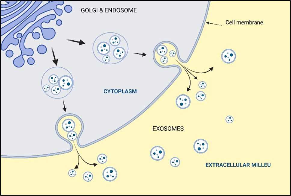 Figure 1. Biogenesis of exosomes (Created in https://BioRender.com).
Among their properties are biocompatibility, stability, low toxicity, and efficient exchange of molecular cargo.⁶ Exosomes can deliver their cargo to specific distant targets to regulate cellular division, survival, differentiation, response to stress, and apoptosis.⁷ Evidence has confirmed that exosomes promote wound healing, stimulate bone regeneration, and enable cartilage repair by inducing angiogenesis, collagen fiber deposition, and inhibiting inflammation.⁸⁻¹⁰ However, due to the technical limitations in obtaining sufficient exosome quantities for clinical applications, no commercial products based on exosomes have been approved by medicine agencies.¹¹ In contrast, autologous-derived exosomes obtained from the patient’s tissues, such as platelets, whole blood, or fat, are emerging as a promising avenue for regenerative treatments.
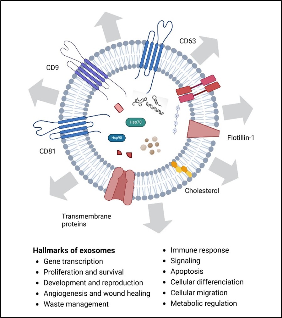 Figure 2. Hallmarks of exosomes (Created in https://BioRender.com).
How can exosomes be characterized, isolated and quantified?
Although emerging detection methods and new analytical techniques exist, there is no purification method to separate exosomes based on their size and no consensus on specific markers that uniquely distinguish the origin of these vesicles once they have left the cell.¹²
Several methods are used to characterize EVs, identify protein composition, and detect nucleic acids and lipids. They include conventional western blotting, mass spectrometry, flow cytometry (bead coupled), microfluidics chips, polymerase chain reaction, and nuclear magnetic resonance (Figure 3). Super-resolving techniques, such as electron microscopy, determine the size, shape, morphology-integrity, inter-particle interaction, and spatial relationship with tissues and cells to complete exosome qualitative information.¹³,¹⁴
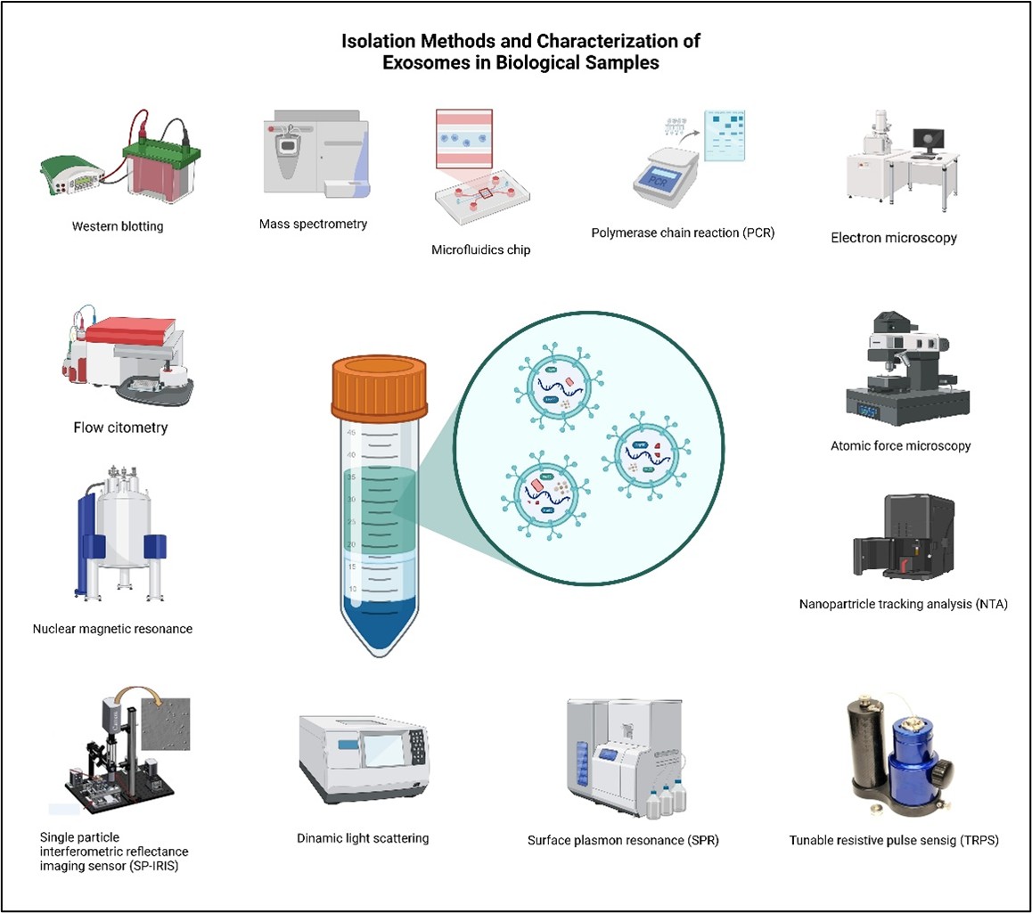
Figure 3. Isolation and characterization methods of exosomes (Created in https://BioRender.com).
Regarding other quantification techniques, we can find atomic force microscopy, nanoparticle tracking analysis (NTA), tunable resistive pulse sensing (TRPS), dynamic light scattering, surface plasmon resonance (SPR), and the single particle interferometric reflectance imaging sensor (SP-IRIS) (Figure 3). Among the isolation techniques are ultracentrifugation, ultrafiltration, size exclusion chromatography, precipitation, and immunoaffinity-based capture. However, each isolation and quantification method have some advantages and disadvantages, particularly regarding the integrity of the exosomes obtained and the specificity of the particles detected.¹⁵,¹⁶
While these issues are solving, treatments with autologous materials, such as platelet-rich plasma or adipose-derived stem cells preconditioned with photothermal biomodulation to stimulate exosome release, have been proven to be efficacious and safe for regenerative medicine.
For more in-depth insights on exosomes, download our free eBook here: https://metacelltech.com/exosomesebook/

References
1. Colombo M, Raposo G, Théry C. Biogenesis, Secretion, and Intercellular Interactions of Exosomes and Other Extracellular Vesicles. Annu Rev Cell Dev Biol [Internet]. 2014 Oct 11;30(1):255–89. Available from: https://www.annualreviews.org/doi/10.1146/annurev-cellbio-101512-122326
2. Meldolesi J. Exosomes and Ectosomes in Intercellular Communication. Current Biology [Internet]. 2018 Apr;28(8):R435–44. Available from: https://linkinghub.elsevier.com/retrieve/pii/S0960982218300927
3. Di Bella MA. Overview and Update on Extracellular Vesicles: Considerations on Exosomes and Their Application in Modern Medicine. Biology (Basel) [Internet]. 2022 May 24;11(6):804. Available from: https://www.mdpi.com/2079-7737/11/6/804
4. Chen H, Wang L, Zeng X, Schwarz H, Nanda HS, Peng X, et al. Exosomes, a New Star for Targeted Delivery. Front Cell Dev Biol [Internet]. 2021 Oct 8;9. Available from: https://www.frontiersin.org/articles/10.3389/fcell.2021.751079/full
5. Tang Y, Zhou Y, Li HJ. Advances in mesenchymal stem cell exosomes: a review. Stem Cell Res Ther [Internet]. 2021;12(1):71. Available from: https://doi.org/10.1186/s13287-021-02138-7
6. Zeng H, Guo S, Ren X, Wu Z, Liu S, Yao X. Current Strategies for Exosome Cargo Loading and Targeting Delivery. Cells [Internet]. 2023 May 17;12(10):1416. Available from: https://www.mdpi.com/2073-4409/12/10/1416
7. Ratajczak MZ, Ratajczak J. Horizontal transfer of RNA and proteins between cells by extracellular microvesicles: 14 years later. Clin Transl Med [Internet]. 2016 Dec 4;5(1). Available from: https://onlinelibrary.wiley.com/doi/10.1186/s40169-016-0087-4
8. Yu L, Qin J, Xing J, Dai Z, Zhang T, Wang F, et al. The mechanisms of exosomes in diabetic foot ulcers healing: a detailed review. J Mol Med [Internet]. 2023 Oct 11;101(10):1209–28. Available from: https://link.springer.com/10.1007/s00109-023-02357-w
9. Littig JPB, Moellmer R, Agrawal DK, Rai V. Future applications of exosomes delivering resolvins and cytokines in facilitating diabetic foot ulcer healing. World J Diabetes [Internet]. 2023 Jan 15;14(1):35–47. Available from: https://www.wjgnet.com/1948-9358/full/v14/i1/35.htm
10. Roszkowski S. Therapeutic potential of mesenchymal stem cell-derived exosomes for regenerative medicine applications. Clin Exp Med [Internet]. 2024 Mar 1;24(1):46. Available from: https://link.springer.com/10.1007/s10238-023-01282-z
11. Asadpour A, Yahaya BH, Bicknell K, Cottrell GS, Widera D. Uncovering the gray zone: mapping the global landscape of direct-to-consumer businesses offering interventions based on secretomes, extracellular vesicles, and exosomes. Stem Cell Res Ther [Internet]. 2023 May 4;14(1):111. Available from: https://stemcellres.biomedcentral.com/articles/10.1186/s13287-023-03335-2
12. Yakubovich EI, Polischouk AG, Evtushenko VI. Principles and Problems of Exosome Isolation from Biological Fluids. Biochem (Mosc) Suppl Ser A Membr Cell Biol [Internet]. 2022 Jun 16;16(2):115–26. Available from: https://link.springer.com/10.1134/S1990747822030096
13. BRYDSON R, BROWN A, HODGES C, ABELLAN P, HONDOW N. Microscopy of nanoparticulate dispersions. J Microsc [Internet]. 2015 Dec 6;260(3):238–47. Available from: https://onlinelibrary.wiley.com/doi/10.1111/jmi.12290
14. Szatanek R, Baj-Krzyworzeka M, Zimoch J, Lekka M, Siedlar M, Baran J. The Methods of Choice for Extracellular Vesicles (EVs) Characterization. Int J Mol Sci [Internet]. 2017 May 29;18(6):1153. Available from: https://www.mdpi.com/1422-0067/18/6/1153
15. Yakubovich EI, Polischouk AG, Evtushenko VI. Principles and Problems of Exosome Isolation from Biological Fluids. Biochem (Mosc) Suppl Ser A Membr Cell Biol [Internet]. 2022 Jun 16;16(2):115–26. Available from: https://link.springer.com/10.1134/S1990747822030096
16. Kurian TK, Banik S, Gopal D, Chakrabarti S, Mazumder N. Elucidating Methods for Isolation and Quantification of Exosomes: A Review. Mol Biotechnol [Internet]. 2021 Apr 25;63(4):249–66. Available from: http://link.springer.com/10.1007/s12033-021-00300-3